
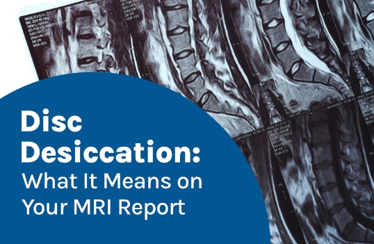
Magnetic resonance imaging (MRI) is a noninvasive diagnostic tool that captures high-resolution 3-dimensional images of an area in question, including soft tissue, bones, and organs. Disc desiccation is part of the degenerative process and contributes to degenerative disc disease (DDD).
Disc desiccation can occur naturally with age. When we’re born, the discs are made largely of water, but over time, they experience fluid loss. Excessive fluid loss is known as desiccation and is a common degenerative change. An MRI report will show a dehydrated disc’s inner nucleus as darker grey, when it should be bright white.
If the spine’s intervertebral discs are degenerating, spinal health can be affected in a number of ways.
Intervertebral Discs
The spine’s intervertebral discs sit between adjacent vertebral bodies and have two main structures: a soft gel-like interior and a tough and flexible outer layer.
The disc’s soft interior is the nucleus pulposus and is made of water, proteoglycans, and collagen fibers. The high water content of the nucleus facilitates its ability to act as the spine’s shock absorbers by evenly absorbing and distributing hydraulic pressure throughout the disc (1).
A disc’s inner water content is estimated at 66 – 86 percent and decreases with age. The proteoglycans attract and hold water molecules, working to keep the disc hydrated (1).
The collagen fibers maintain the disc’s structure and facilitate its strength.
A disc’s outer layer is known as the annulus fibrosus and contains layers of collagen fibers capable of resisting tension and compressive force (excessive and/or uneven pressure) while maintaining the central position of the nucleus inside the disc (1).
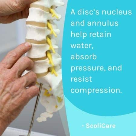
A disc’s inner nucleus is flexible but durable and works with the outer annulus to maintain its water content and ability to absorb and distribute hydraulic pressure and resist compressive force (1).
Disc Desiccation on an MRI Report
So what does disc desiccation mean for disc and spinal health? Disc desiccation can occur slowly over time as the discs experience a decrease in water content.
On an MRI, a hydrated and healthy disc will appear bright white due to its high water content, and a dehydrated/degenerating disc will appear darker in color and is also likely to show a loss of height (2).
If a disc is bulging or herniated, it will also show on an MRI (2).
Disc Desiccation
When the spine experiences degenerative changes, it often starts with the discs losing hydration and changing shape as a result (3).
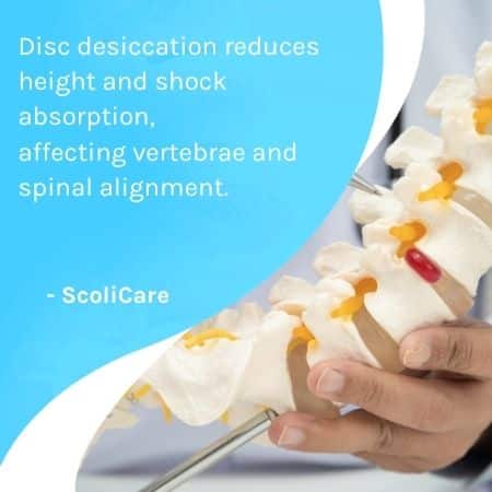
With desiccation, a disc loses height, becomes thinner, more rigid, and less able to absorb shock, and this also affects adjacent vertebrae attached and their alignment with the rest of the spine (3).
Degenerative disc disease contributes to a number of spinal conditions and issues, making the spine more vulnerable to injury and strain.
Disc desiccation takes place slowly over time and because the discs are largely avascular, they are slow to repair and heal. The discs need to absorb oxygen and nutrients needed for repair through diffusion (1, 3).
Once a certain level of disc desiccation has occurred, discs are at risk of becoming bulging and/or herniated.
Bulging and Herniated Discs
A bulging disc develops when the inner nucleus is pushing outwards against its outer annulus, causing the disc to bulge into its surroundings, and if a disc’s inner nucleus pulposus pushes through a tear in the outer layer, it has herniated (3).
Disc desiccation starts the degenerative cycle that once initiated, is difficult to slow and/or reverse (3).
A dehydrated disc will lose cushioning and height, disrupting its ability to prevent friction between adjacent vertebrae and provide structural support for the spine’s alignment (1, 3).
Excessive loss of water inside the disc causes the outer layer to lose elasticity, and the more brittle it becomes, the more vulnerable it is to tears, herniation, and further damage (3).
In many cases, desiccation is the start of degenerative disc disease (3).
Degenerative Disc Disease Risk Factors
A certain level of degeneration is expected with age, but there are lifestyle factors that can shape spinal health over time and accelerate degenerative changes, often impacting discs first.
Obesity is a common cause of disc and back issues due to the extra weight and stress it puts on the spine and its structures (4).
Low activity levels can also contribute to disc desiccation over time by depriving the discs of pressure changes through movement that promotes healthy blood flow, circulation, and the diffusion of nutrients needed for repair and eliminating waste (1, 3).
Chronic poor posture can also shape spinal health over time by exposing the spine and its supportive muscles and ligaments to uneven forces and strain (5).
Smoking and excessive consumption of alcohol can also contribute to disc degeneration by reducing blood flow (6).
Occupation can also play a role if it includes repeated stress from heavy lifting and strenuous movements that strain the vertebrae and discs of the lumbar spine (7).
Symptoms of Disc Desiccation
Symptoms of disc desiccation develop from a loss of the disc’s cushioning, and common symptoms include chronic localized back around the affected disc, along with stiffness and a reduced range of motion (3).
When a disc changes shape, it can take up extra space within the spine used for the spinal nerves to function optimally within, and nerve compression can cause pain, tingling, weakness, and/or numbness in the arms and legs (3).
In severe cases, it can disrupt bowel and bladder functions (3).
Disc Desiccation Treatment Options
The first line of treatment options for a desiccated disc is conservative, focusing on symptom relief, preventing further degeneration, and improving disc function.
Physical therapy works towards increasing core strength for more spinal support and less pressure on its individual structures and discs. A strong core is needed to maintain healthy and upright posture (8).
Physical therapy also targets the spine’s flexibility to improve range of motion and mobility.
Healthy movement is key to disc health as the discs receive nutrients and oxygen needed for repair through diffusion, so increasing blood flow and circulation in the area can improve disc health and function (8).
Medication for pain relief and reducing inflammation may help with symptom relief, and lifestyle changes can include weight loss for less pressure on the spinal discs (8).
Education on the ergonomics of heavy lifting can help prevent further damage (8).
Spinal decompression therapy can also be applied to create space within the spine, taking pressure off the discs and improving blood flow (8).
If conservative treatment options are unsuccessful, severe cases involving chronic pain and/or neurological symptoms may require surgical intervention (laminectomy, discectomy, or spinal fusion) (8).
Conclusion
There are a number of factors that shape overall spinal health, and disc health and function is paramount.
The discs combine forces to facilitate the spine’s flexibility, and because adjacent vertebrae attach to the disc in between, they provide the spine with crucial structural support, a loss of which can cause the spine to shift out of alignment.
The discs also act as cushions to prevent friction during movement and help with shock absorption.
If a disc becomes dehydrated (a common effect of aging), it can lose height, become thinner, more brittle, and less functional.
Disruptions to disc function can alter the spine’s alignment, strength, flexibility, and ability to absorb shock.
Disc desiccation develops slowly over time and common symptoms include localized back pain and/or pain that radiates into the arms and legs due to nerve compression and reduced mobility.
Common treatment options for preventing further degeneration and improving the function of a degenerating disc include physical therapy, medication, chiropractic care, and lifestyle improvement, and in severe cases, surgical intervention may be recommended.
References:
- Waxenbaum JA, Reddy V, Futterman B. Anatomy, Back, Intervertebral Discs. [Updated 2023 Dec 9]. In: StatPearls [Internet]. Treasure Island (FL): StatPearls Publishing; 2025 Jan-. Available from: https://www.ncbi.nlm.nih.gov/books/NBK470583/
- Suthar P, Patel R, Mehta C, Patel N. MRI evaluation of lumbar disc degenerative disease. J Clin Diagn Res. 2015 Apr;9(4):TC04-9. doi: 10.7860/JCDR/2015/11927.5761. Epub 2015 Apr 1. PMID: 26023617; PMCID: PMC443713
- Scarcia L, Pileggi M, Camilli A, Romi A, Bartolo A, Giubbolini F, Valente I, Garignano G, D’Argento F, Pedicelli A, Alexandre AM. Degenerative Disc Disease of the Spine: From Anatomy to Pathophysiology and Radiological Appearance, with Morphological and Functional Considerations. J Pers Med. 2022 Nov 1;12(11):1810. doi: 10.3390/jpm12111810. PMID: 36579533; PMCID: PMC9698646
- Samartzis D, Karppinen J, Cheung JP, Lotz J. Disk degeneration and low back pain: are they fat-related conditions? Global Spine J. 2013 Jun;3(3):133-44. doi: 10.1055/s-0033-1350054. Epub 2013 Jul 17. PMID: 24436864; PMCID: PMC3854598
- Fasser MR, Furrer PR, Fisler L, Urbanschitz L, Snedeker JG, Farshad M, Widmer J. The triadic relationship between spinal posture, loading, and degeneration. Front Bioeng Biotechnol. 2025 Mar 18;13:1444540. doi: 10.3389/fbioe.2025.1444540. PMID: 40171041; PMCID: PMC11959076
- Rajesh N, Moudgil-Joshi J, Kaliaperumal C. Smoking and degenerative spinal disease: A systematic review. Brain Spine. 2022 Aug 7;2:100916. doi: 10.1016/j.bas.2022.100916. PMID: 36248118; PMCID: PMC9560562
- Pope MH, Goh KL, Magnusson ML. Spine ergonomics. Annu Rev Biomed Eng. 2002;4:49-68. doi: 10.1146/annurev.bioeng.4.092101.122107. Epub 2002 Mar 22. PMID: 12117750
- Donnally III CJ, Hanna A, Varacallo MA. Lumbar Degenerative Disk Disease. [Updated 2023 Aug 4]. In: StatPearls [Internet]. Treasure Island (FL): StatPearls Publishing; 2025 Jan-. Available from: https://www.ncbi.nlm.nih.gov/books/NBK448134/
More news
-
 21 August 2026
Multilevel degenerative disc disease affects discs in multiple spinal areas, impacting flexibility, stability, and movement. Early care can help manage symptoms.
-
 3 February 2026
Early detection and customized nonsurgical care—like 3D bracing and scoliosis-specific rehab—can help stop progression and improve spinal health.
-
 6 February 2024
As students head back to school, a trusty backpack becomes an essential companion. However, the convenience of carrying books, laptops, and supplies on your back comes with a responsibility to…