
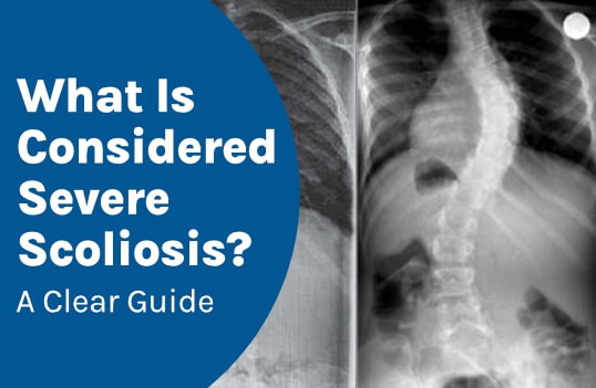
Scoliosis severity is determined by curve size and treatment plans are crafted in response. A number of measurements are taken during a patient’s initial scoliosis X-ray; their accuracy is crucial. Severe scoliosis involves larger curves that are more complex to treat.
Scoliosis severity ranges from mild to severe and is based on the size of the unnatural spinal curve. While there are additional factors that shape a patient’s scoliosis severity and rate of progression, a patient’s Cobb angle measurement classifies scoliosis as mild, moderate, or severe.
Severe scoliosis requires a more comprehensive treatment plan than mild cases.
Severe Scoliosis
Severe scoliosis involves larger curves, and treatment plans are shaped around curve size and severity.
Because scoliosis is progressive, even cases initially diagnosed as mild or moderate scoliosis can change over time, likely to become severe if left untreated, but scoliosis can be highly treatable, when plans are proactive and work towards preventing mild cases from becoming severe (1).
The main trigger for progression is growth, so cases of childhood scoliosis can involve rapid scoliosis progression. Adolescent idiopathic scoliosis is the most prevalent form, and due to the rapid nature of pubescent growth spurts, adolescent scoliosis can become severe quickly (1, 2).
The goal with every patient is early detection and intervention. No treatment outcome can be guaranteed, but particularly in cases of childhood scoliosis, early intervention is key, and treatment plans are customized around important variables such as severity (1, 2).
Severe scoliosis is more complex to treat than mild scoliosis and moderate cases, so the goal is to keep mild cases mild and prevent further curve progression and increasing symptoms (1).
Severe Scoliosis Symptoms
Severe scoliosis symptoms can be overt (3). Scoliosis can be pre-existing or develop fresh in adulthood as degenerative de Novo scoliosis (4).
Scoliosis affects all ages and most commonly develops during adolescence as adolescent idiopathic scoliosis, and the main symptoms of severe childhood scoliosis are overt postural and mobility changes (2).
The larger the size of the spine’s unnatural lateral spinal curve, the more severe the scoliosis, and the more noticeable symptoms are going to be. Another benefit to treating scoliosis proactively and working towards preventing progression is minimizing the potential effects and complications of severe scoliosis (1).
While there are a number of factors that shape a patient’s experience of scoliosis and treatment needs, determining condition severity accurately is an important first step towards a positive treatment outcome.
How is Scoliosis Severity Determined?
A measurement known as Cobb angle assesses the degree of scoliosis, and an X-ray is needed to measure a patient’s Cobb angle, assess, and diagnose scoliosis (5).
Cobb angle measurement is the most widely-used method for assessing scoliosis. Smaller mild curves are measured at less than 20 degrees, and curves more than 35 degrees are diagnosed as moderate scoliosis; curves measuring more than 60 degrees are considered severe (5).
The higher a Cobb angle, the more severe the scoliosis, and the more complex it can be to treat (5). The benefit of diagnosing and treating scoliosis with early intervention is the potential to prevent mild and moderate curves from becoming severe (1).
Cobb Angle Measurement Accuracy
A patient’s Cobb angle is a key piece of information used to quantify curve magnitude and guide treatment plans, so it needs to be as precise and accurate a measurement as possible (5).
Determining a Cobb angle involves locating the most-tilted vertebrae of the curve. Lines are drawn from the endplates of the most misaligned vertebrae, and the intersecting angle is represented in degrees (5).
Many treatment decisions are based on comparisons between a patient’s Cobb angle at the time of diagnosis and how/if it changes throughout the course of treatment; in childhood scoliosis treatment, there is also a focus on how a patient’s Cobb angle measurement responds to growth (5, 6).
In addition to Cobb angle, the angle of trunk rotation, curve location, scoliosis type, and patient age are additional key factors shaping the course of treatment, and the results of a patient’s initial scoliosis X-ray provides most of this information (5, 7).
Scoliosis X-Ray
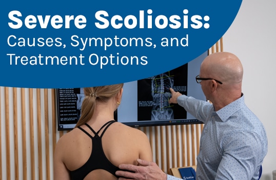
X-ray imaging is needed to diagnose scoliosis, determine its severity, the degree of rotation, where the curve is located, predict progression, and assess overall spinal health and function (7).
When scoliosis is severe, a large curve is involved, and scoliosis that develops in the thoracic spine is the most likely to progress quickly and become severe (8).
In older adults, the spine’s balance and stability need to be accurately assessed for fall-prevention purposes, and in children, X-rays can also assess bone maturity to predict the rate of curve progression (9, 10).
Scoliosis isn’t static; it changes over time. As a progressive condition triggered by growth, a customized treatment plan needs to allow for adjustments based on how a patient’s curve is responding to treatment and/or growth (6).
Patients should be aware that determining a Cobb angle measurement isn’t a simple process, and even a minor variance of a degree is significant, when it comes to customizing treatment plans for precise results (5).
Standing X-rays are recommended for accuracy as the curve flattens out when the patient is lying down, and it’s also important to measure the spine while it’s supporting body weight and subject to gravitational pressure (5).
So severe scoliosis cases involve large curves that commonly involve overt symptoms, and while the goal is always early diagnosis and intervention, all severity levels require treatment (1).
Severe Scoliosis Treatment
Correcting severe scoliosis can be challenging due to spinal rigidity; as scoliosis progresses, the spine becomes increasingly rigid; this is a primary benefit of starting treatment early, when the spine’s position can be more-easily improved and corrected (11).
Severe scoliosis cases can involve patients with typical types, atypical and rare types, patients whose scoliosis has progressed slowly over time, patients whose scoliosis has progressed rapidly, and cases that have developed later in life due to degenerative instability (2, 4, 12).
Most cases of scoliosis involve idiopathic scoliosis, considered typical, and less-common types are associated with known causes such as neuromuscular scoliosis caused by the presence of a neuromuscular condition like cerebral palsy; these cases can be among the most severe and complex to treat because the neuromuscular condition has to be addressed to impact the scoliosis (12).
Most cases of adult scoliosis involve idiopathic scoliosis that’s pre-existing from adolescence, and older adults can develop degenerative de novo scoliosis later in life, and because degenerative scoliosis is caused by degenerative instability, most cases are progressive and painful (4, 9).
Treatment for adult scoliosis focuses on increasing the spine’s alignment, balance, and stability for fall prevention and pain relief (4).
Treatment for childhood scoliosis focuses on counteracting progression during growth through curvature reductions (6).
Nonsurgical Treatment
While milder cases may respond to scoliosis-specific rehabilitative exercise and/or the prescription of part-time corrective bracing, with moderate scoliosis curves more than 35 degree, full-time corrective bracing is often favored as the primary form of treatment over scoliosis-specific exercise alone (13).
ScoliBalance® is a customized scoliosis-specific rehabilitative exercise program that facilitates 3-dimensional correction through improvements to curve size, posture, spinal flexibility, and targeting the spine’s surrounding muscle balance and strength for more support (13).
A ScoliBrace® can further support 3-dimensional improvements by maintaining the spine and body’s straight and upright posture (13).
For severe curves of more than 60 degrees, surgical treatment may be necessary, and this most often involves spinal fusion surgery that realigns the spine and holds it in place with attached rods (11).
A goal of proactive nonsurgical treatment options is to help patients avoid the need for surgical intervention (1).
Conclusion
When scoliosis is diagnosed early and treated proactively through scoliosis-specific physical therapy and corrective bracing, the potential for a successful nonsurgical treatment outcome is more likely.
A diagnosis of mild scoliosis doesn’t mean it will stay mild, not without the help of a proactive customized treatment plan.
As a progressive spinal condition, treatment guarantees can’t be given, and treatment needs are ongoing, but many cases of scoliosis are highly treatable, and the sooner treatment is started, and the more customized it is, the better.
The best way to minimize the potential effects and complications of scoliosis is by preventing mild and moderate cases from becoming severe.
References:
- McCarthy RE. Prevention of the complications of scoliosis by early detection. Clin Orthop Relat Res. 1987 Sep;(222):73-8. PMID: 2957138.
- Weinstein SL. The Natural History of Adolescent Idiopathic Scoliosis. J Pediatr Orthop. 2019 Jul;39(Issue 6, Supplement 1 Suppl 1):S44-S46. doi: 10.1097/BPO.0000000000001350. PMID: 31169647
- Janicki JA, Alman B. Scoliosis: Review of diagnosis and treatment. Paediatr Child Health. 2007 Nov;12(9):771-6. doi: 10.1093/pch/12.9.771. PMID: 19030463; PMCID: PMC2532872
- Aebi M. The adult scoliosis. Eur Spine J. 2005 Dec;14(10):925-48. doi: 10.1007/s00586-005-1053-9. Epub 2005 Nov 18. PMID: 16328223
- Horng MH, Kuok CP, Fu MJ, Lin CJ, Sun YN. Cobb Angle Measurement of Spine from X-Ray Images Using Convolutional Neural Network. Comput Math Methods Med. 2019 Feb 19;2019:6357171. doi: 10.1155/2019/6357171. PMID: 30996731; PMCID: PMC6399566
- Negrini, S., Donzelli, S., Aulisa, A. G., Czaprowski, D., Schreiber, S., de Mauroy, J. C., … & Zaina, F. (2018). 2016 SOSORT guidelines: orthopaedic and rehabilitation treatment of idiopathic scoliosis during growth. Scoliosis and spinal disorders, 13(1), 3
- Kim H, Kim HS, Moon ES, Yoon CS, Chung TS, Song HT, Suh JS, Lee YH, Kim S. Scoliosis imaging: what radiologists should know. Radiographics. 2010 Nov;30(7):1823-42. doi: 10.1148/rg.307105061. Erratum in: Radiographics. 2015 Jul-Aug;35(4):1316. doi: 10.1148/rg.2015154011. PMID: 21057122
- Wong LPK, Cheung PWH, Cheung JPY. Curve type, flexibility, correction, and rotation are predictors of curve progression in patients with adolescent idiopathic scoliosis undergoing conservative treatment : a systematic review. Bone Joint J. 2022 Apr;104-B(4):424-432. doi: 10.1302/0301-620X.104B4.BJJ-2021-1677.R1. PMID: 35360948; PMCID: PMC9020521
- Kotwal S, Pumberger M, Hughes A, Girardi F. Degenerative scoliosis: a review. HSS J. 2011 Oct;7(3):257-64. doi: 10.1007/s11420-011-9204-5. Epub 2011 Jun 11. PMID: 23024623; PMCID: PMC3192887
- Andleeb I, Hussain BZ, Joncas J, Barchi S, Roy-Beaudry M, Parent S, Grimard G, Labelle H, Duong L. Automatic Evaluation of Bone Age Using Hand Radiographs and Pancorporal Radiographs in Adolescent Idiopathic Scoliosis. Diagnostics (Basel). 2025 Feb 13;15(4):452. doi: 10.3390/diagnostics15040452. PMID: 40002603; PMCID: PMC11854906
- Gatam L, Phedy P, Mahadhipta H, Husin S, Gatam AR, Kadar PD, Gani KS, Mitchel M, Kholinne E. Surgical outcomes and complication rates in severe scoliosis: a systematic review. SICOT J. 2025;11:53. doi: 10.1051/sicotj/2025050. Epub 2025 Sep 19. PMID: 40971604; PMCID: PMC12448635
- Vialle R, Thévenin-Lemoine C, Mary P. Neuromuscular scoliosis. Orthop Traumatol Surg Res. 2013 Feb;99(1 Suppl):S124-39. doi: 10.1016/j.otsr.2012.11.002. Epub 2013 Jan 19. PMID: 23337438
- Marchese R, Du Plessis J, Pooke T, McAviney J. The Improvement of Trunk Muscle Endurance in Adolescents with Idiopathic Scoliosis Treated with ScoliBrace® and the ScoliBalance® Exercise Approach. J Clin Med. 2024 Jan 23;13(3):653. doi: 10.3390/jcm13030653. PMID: 38337346; PMCID: PMC10856658
More news
-
 8 August 2025
What patients need to understand about severe scoliosis is that it doesn't start that way; as a progressive condition, scoliosis severity can increase over time, and while young patients can…
-
 15 July 2024
In recent years, patient-centred care has revolutionised the approach to scoliosis treatment by focusing on the unique needs, preferences and values of each patient. This approach places patients at the…
-
 24 February 2023
The short answer is yes, people with scoliosis can live normally. However, it is important to understand the condition and its impact on the individual's life. Scoliosis is a complex…