Case – Adult Bracing (61 year old female)
Managing Adolescent Idiopathic Scoliosis in the Adult with ScoliBrace® and ScoliBalance®
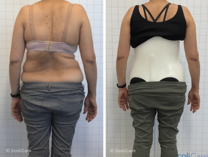
At 61 years old, the patient visited the ScoliCare clinic after experiencing a deterioration in posture,pain and an inability to stand or walk for long periods of time. An X-ray taken prior to attending ScoliCare revealed a scoliosis measurement of 49°.
Within 10 years, her scoliosis progressed to 59° and her function ability declined without treatment. This patient was diagnosed with Adolescent Idiopathic Scoliosis in the Adult, with degenerative changes. The patient was prescribed a part-time ScoliBrace® and ScoliBalance® treatment plan to manage the progression and symptoms.

Scoliosis Stabilized: After 6 years of ScoliBrace® and ScoliBalance®, her scoliosis that was aggressively progressing, remains stable at 59°, with no further progression.
Improved Posture: The patient has noticed a significant change in her posture and is more confident.
Improved Function and Comfort: The patient experienced a significant reduction in pain and an increased ability to stand and walk for extended periods.